Spontaneous vs. directed self-organization (DSO)
Nature exhibits many ordered or quasi-ordered self-organized patterns, such as parallel stripes on sand dunes, sea shells, and animal skins, as well as the convoluted structures of human brains and proteins (Fig.1a). Self-organization (SO) is also ubiquitous in chemistry, material science, and biology, and can involve objects ranging from molecular to centimeter scales (Whitesides and Boncheva, PNAS 99(8), p. 4769-4774, 2002). Nature utilizes SO to produce extremely large numbers of small scale structures at a rapid rate, such as the formation of molecular crystals, colloids, lipid bilayers, phase-separated polymers, self-assembled monolayers, as well as folding of proteins and DNA. According to Gier-Meinhardt model, pattern formation results from competition between positively- and negatively- feedback processes (http://www.scholarpedia.org/article/Gierer-Meinhardt_model). These interactions are weak and local in nature, leading to many degeneracies in configuration space and a lack of long-range order in patterns. This explains why zebras all have stripes but none has identical patterns! Another example is the block copolymers, which under normal condition form swirling patterns due to microphase separation (Fig.1b). To increase long-range order, external stimuli are needed to break degeneracy to direct self-organization in desired ways. For example, Kim and co-workers have shown that chemically altering the surface using lithography directs the self-organization of block copolymers into straight and parallel lines (Kim, et al., Nature, 2003. 424(6947): p. 411-414). In parallel, directed self-organization (DSO) of nanoparticles (NPs) has also received strong interests. Learning the rules for consistently arranging NPs, like nature arranges atoms into molecules and materials, is essential to capitalize on nanotechnology’s potential for broad application. For example, Mirkin and co-workers at Northwestern University has recently reported using DNA as molecular linkers to assemble gold NPs into crystalline lattices with design specific configurations (Fig.1c) (NSF press release 11-218). The other prominent example is the formation of ordered quantum dots using strain engineering of substrates (Fig.1d) (Shiryaev, et al., Thin Solid Films, 1997. 294(1-2): p. 311-314).
Figure 1: (a) Self-organized patterns in nature: sand dunes, sea shells, zebra skins, human brains, and proteins. (b) DSO of a block copolymer film. (c) Assembly of gold NPs crystals using DNA linkers. (d) DSO of quantum dots. Images sources: sand dune (http://www.math.ubc.ca/~conway/images/tb_mui_ne_sand_dunes.jpg), zebra (http://www.hdpicturesofanimals.com/hd-animals-pictures/zebra-8.jpg), sea shells (http://www.scholarpedia.org/article/File:Self_organization_10.jpg), brain (brain.jpg), protein (http://techglimpse.com/wp-content/uploads/2009/01/p1-300×300.jpg), block copolymers (Kim, et al., Nature, 2003. 424(6947), p. 411-414), gold NPs DSO (NSF press release 11-218), ASQD (Shiryaev, et al., Thin Solid Films, 1997. 294(1-2): p. 311-314).Femtosecond-laser-induced periodic self-organized nanostructures (FLIPSON)
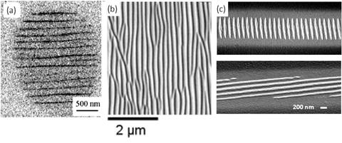
It is well known that femtosecond-pulsed (fs) laser, by localizing its energy in space (i.e., tightly focused) and time (i.e., short pulse duration), can create confined nanostructures (e.g., holes, voids, etc) with dimension less than 100 nm. Surprisingly, fs laser can also achieve nanostructuring without energy localization: within the smooth laser intensity profile, periodic nanostructures form spontaneously without any beam shaping. This phenomenon, dubbed femtosecond laser-induced periodic self-organized nanostructures (FLIPSON), has been observed in many different experimental settings, including nanocracks inside fused silica, nanogratings on dielectric surface by ablation, and on dielectric surface by deposition (Fig. 2). Generally speaking, FLIPSON is produced over many laser shots with pulse energy lower than the single-shot damage threshold and is characterized by a deep sub-wavelength period that is smaller than half of the light wavelength (L £ l/2), both of which make FLIPSON drastically different from the classical rippling phenomena known as LIPSS (van Driel et. al., PRL 1982, 49(26), p. 1955-1958). Understanding FLIPSON will enrich our knowledge of light-matter interaction at femtosecond time and nanometer length scales, which could have significant technical impacts. On one hand, FLIPSON might represent unwanted defects for femtosecond-laser material processing and removing it could help achieve better work quality. On the other hand, FLISPON presents a rich opportunity in nanoscience and engineering to enable alternative techniques and principles for cost-effective and large-scale nanopatterning.
For a comprehensive review on this subject, please refer to my book chapter (Her, chapter 4.10, Comprehensive Nanoscience and Technology, edited by David Andrews, Greg Scholes, and Gary Wiederrecht, Elsevier 2011).
Figure 2: Examples of FLIPSON: (a) nanocracks inside fused silica (Shimotsuma et. al., PRL 91, p. 247405, 2003); (b) nanogratings on surface of dielectrics by ablation (Reif et. al., APA 92, pp. 1019-1024, 2008); (c) tungsten nanogratings on dielectrics by deposition (Zhang et. al., Optics Express 15, pp. 5937-5947, 2007).Tungsten nanograting (TNG) – An example of FLIPSON observed during laser CVD of tungsten
Our unique contribution in this field is the discovery of tungsten nanogratings (TNGs). This observation came un-expected and is a god example of serendipity (http://en.wikipedia.org/wiki/Serendipity#Role_in_science_and_technology). The quality of some TNGs can be so good that it reminds us the textbook example of interference fringes of two coherent laser beams by Thompson and Wolf in 1957 (Fig. 3 (a)), except our TNG is made by a single laser beam! Like many other examples of serendipity, we truly hope our observation of nanogratings, or more generally, self-organized periodic nanostructures formed during LCVD, can turn useful to the mankind. Current we are engaged in mechanistic study of TNGs where we have many mysterious results to be explained. As an example, Fig. 3 (b) shows the evolution of TNGs from low to high photon does, indicating that TNG is seeded by nanoparticles that are self-organized into periodic fashion, followed by concatenation of nanoparticles along the laser polarization to form rods.
Figure 3: (a) Comparison and contrast between 2-beam interference fringe and TNG. (b) Evolution of steady-state TNG pattern vs. laser scanning speeds for vertical light polarizations.Current Accomplishments
Reports of tungsten nanogratings (TNG) induced femtosecond laser
1. H. Zhang, M. Tang, J. McCoy, and T. Her, “Deposition of tungsten nanogratings induced by a single femtosecond laser beam,” Optics Express 15 5937 (2007).
2. Mingzhen Tang, Haitao Zhang, and Tsing-Hua Her, “Self-assembly of tunable and highly-uniform tungsten nanogratings induced by femtosecond laser with nanojoule energy,” Nanotechnology 18 (2007) 485304 (5pp).
A comprehensive and updated review of FLIPSON, including FLIPSS inside fused silica, FLIPSS on the surface of dielectrics, and growth of TNGs on surface of dielectrics.
3. Tsing-Hua Her, “Femtosecond-Laser-Induced Periodic Self-Organized Nanostructures,” appeared in Comprehensive Nanoscience and Technology, edited by David Andrews, Greg Scholes, and Gary Wiederrecht, published by Elsevier (Dec. 2010).
Using TNG as patterned substrates for selective growth of tungsten oxides via vapor-solid process.
4. Haitao Zhang, Terry T. Xu, Mingzheng Tang, Tsing-Hua Her, and Shu-you Li, “Selective growth of tungsten oxide nanowires via a vapor-solid process,” J. Vacuum Science and Technology B 28 (2), pp. 310-315 (2010).
Mechanistic study of TNG.
5. Many very interesting results are not yet fully analyzed.
Funding acknowledgements
This project gratefully acknowledges the financial support from DRAPA (#W911NF-05-2-0053) and NSF (#0708555).


